

Immunopathology Core
About
The Immunopathology Core (IPC) provides state-of-the-art immunology and pathology services to COBRE, OCRID, OSU/Oklahoma, and external investigators. The IPC provides advanced immunology and pathology techniques, instrumentation, and expertise required for complex analyses. This core facility serves as a centralized resource offering technical services essential for advancing research primarily in respiratory and infectious diseases. In addition, the IPC collaborates with other OCRID cores, such as the Animal Models Core (AMC) and Molecular Biology Core (MBC) to provide streamlined services to users. The IPC is staffed with an expert immunologist and a board-certified veterinary pathologist each with extensive expertise in animal models and respiratory/infectious disease research. Additional support also includes an experienced core manager who is well trained in advanced immunology and pathology techniques and a histotechnologist to provide additional technical support and enhance service delivery.
COBRE funding in Phases I-II facilitated the establishment, expansion, and enhancement of IPC equipment, personnel, and services. The goals of the IPC during the current Phase III COBRE funding are focused on developing the core into a self-sustaining entity by providing state-of-the-art equipment use to OCRID, Oklahoma, and external investigators, including those from other veterinary colleges.
OCRID Core Facilities Service Request Form
Please use the online form linked below to request a service from our core facility. Thank you!
-
People
Director:
 Rudra Channappanavar, BVSc, PhD.
Dr. Channappanavar is a viral immunologist with 15 years of experience studying immunology of infectious diseases. He has extensive expertise in flow cytometry/immunophenotyping, multiplex cytokine assays, ELISA, immunofluorescence, immunohistochemistry, and serological assays.Co-Director:
Sunil Nivrutti More, BVSc & AH, MVSc, PhD, Diplomate ACVP
Dr. Sunil More is a board-certified veterinary anatomic pathologist specializing in respiratory pathology and host–pathogen interactions. His research focuses on viral respiratory infections and their bacterial complications, with particular emphasis on disease mechanisms and pathogenesis. He has extensive expertise in developing respiratory disease models and lesion characterization with quantitative lesion scoring, providing rigorous pathological endpoints for experimental studies.
Laboratory Manager:
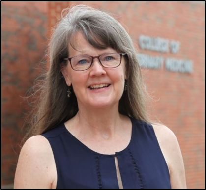 Ms. Shannon Cowan, BS. Ms. Cowan is a skilled in multi parametric flow cytometry and multiplex cytokine assays including prep, collection and analysis. She is a BD trained flow cytometry expert and a cross trained expert in histotechnology and immunohistochemistry. Ms. Cowan manages day-to-day IPC activities and is the primary contact for IPC related questions.
Histotechnologist:
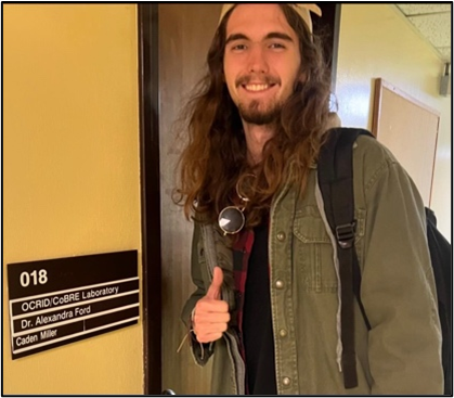 Mr. Caden Miller. Caden is an undergraduate student working as a part-time histotechnician in IPC for the last 3 years.
 -
Contact Information
The Immunopathology Core
171 McElroy Hall
Stillwater, 74078
Phone: 405-744-4749
Email: immunopathologycore@okstate.eduReach out to us! Our staff and directors are always eager to discuss how we can best facilitate your research needs.
-
Histology Fee Sheet
Immunopathology Core: Histology
Service
Unit Charge
1.
Histopathologic evaluation
$120/hour
2.
Gross necropsy eval, tissue collection, and histopathology
$150/hour
3.
Tissue trimming only
$6/cassette
4.
Processing fixed tissue and paraffin embedding only
$8/block
5.
Unstained slides or scrolls from tissue or existing block
$10/slide
6.
Trimming, processing and H&E or special stain slide prep (no interpretation)
$13/slide
7.
Special stains from paraffin embedded tissue
$8/slide
8.
Research IHC*
$50/slide
*Cost: may increase with optimization, labor, cost of primary antibody, purchase of antibody, etc. Adjusted as needed
9.
Recuts from paraffin embedded tissue**
$10/slide
**Cost: may increase with labor, depth required, attempts, etc. Adjusted as needed.
10.
Slide scanning
$5/slide
11.
Trimming, processing, and H&E slide prep (non-research, with interpretation)
$50/animal (Up to 4 slides)
12.
Cryostat (unassisted)
$15/hour
13.
Cryostat (assisted)
$45/hour
14.
Large slide box
$14/box
15.
Small slide box
$8/box
16.
Technical support including training, consultation, IHC/special stain optimization, etc.
$60/hour
The listed prices are for academic users. Active CoBRE pilot projects will be charged 75% of the fee schedule and non-academic users will be charged 1.496 of the fee schedule. b) Project-specific consumables and reagents will be provided by or additionally charged to the investigators.
-
Immunology Fee Sheet
Immunopathology Core: Immunology
Service
Unit Charge
FACS Aria with Operator
$60/hour
FACS Aria self-service (only well-trained personnel)
$30/hour
FACS Aria- Cell Sorting
$80/hour
Cytek Aurora Evo Spectral flow cytometer (Self-service)
$30/hour
Cytek Aurora Evo Spectral flow cytometer (Assisted service)
$60/hour
Flow Cytometry- Data Analysis
$30/hour
Bio-Plex Immunoassay
$45/plate
Bio-Plex Immunoassay- Data Analysis
$30/plate
NanoCellect WOLF cell sorter
$45/hour
Biotek Cytation 5 (Self-Service)
$15/hour
Biotek Cytation 5 (Assisted service)
$45/hour
Biotek Cytation 10 (Self-Service)
$30/hour
Biotek Cytation 10 (Assisted service)
$60/hour
Zeiss 900 LSM (Self-Service)
$30/hour
Zeiss 900 LSM (Assisted service)
$60/hour
Dispens Cell (Self-Service)
$30/hour
Dispens Cell (Assisted service)
$60/hour
The listed prices are for academic users. Active CoBRE pilot projects will be charged 75% of the fee schedule and non-academic users will be charged 1.496 of the fee schedule. b) Project-specific consumables and reagents will be provided by or additionally charged to the investigators.
-
Equipment
FACSAria Special Order Research Product: The Aria has 4 lasers (violet, blue,
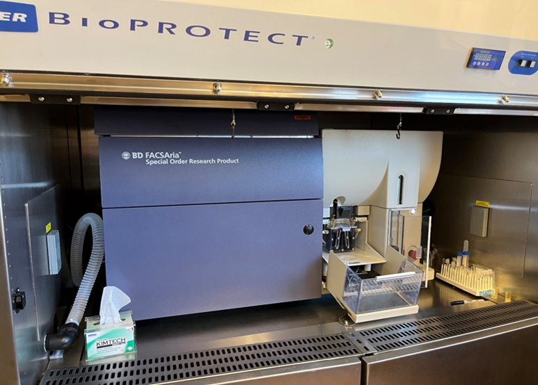 yellow-green, red) with 13 fluorescence parameters for phenotyping. It is capable of aseptic, 4-way (4 cell types) sorts. The automated cell deposition unit (ACDU) allows sorting into multiwell plates and on microscope slides. The cytometer is housed in a Baker BioPROTECT IV biosafety cabinet to allow sorting of BSL2 infected cells or pathogens.
For assistance in designing a panel please contact Ms. Shannon Cowan or refer to this SITE.Flow Cytometry Appointment Policy
Please cancel your flow cytometry appointments as soon as you are aware that you will not be able to attend. Cancellations made less than one hour before the scheduled time will incur a charge equivalent to one hour of instrument use. Failure to show up for a scheduled appointment will also result in a one-hour instrument charge. Please note that billing is based on the scheduled start time of your appointment, not the actual time of arrival.Best Practices for Flow Cytometry
Best Practices for Flow Cytometry2Cytek Aurora Evo: The Cytek Aurora EVO is an advance 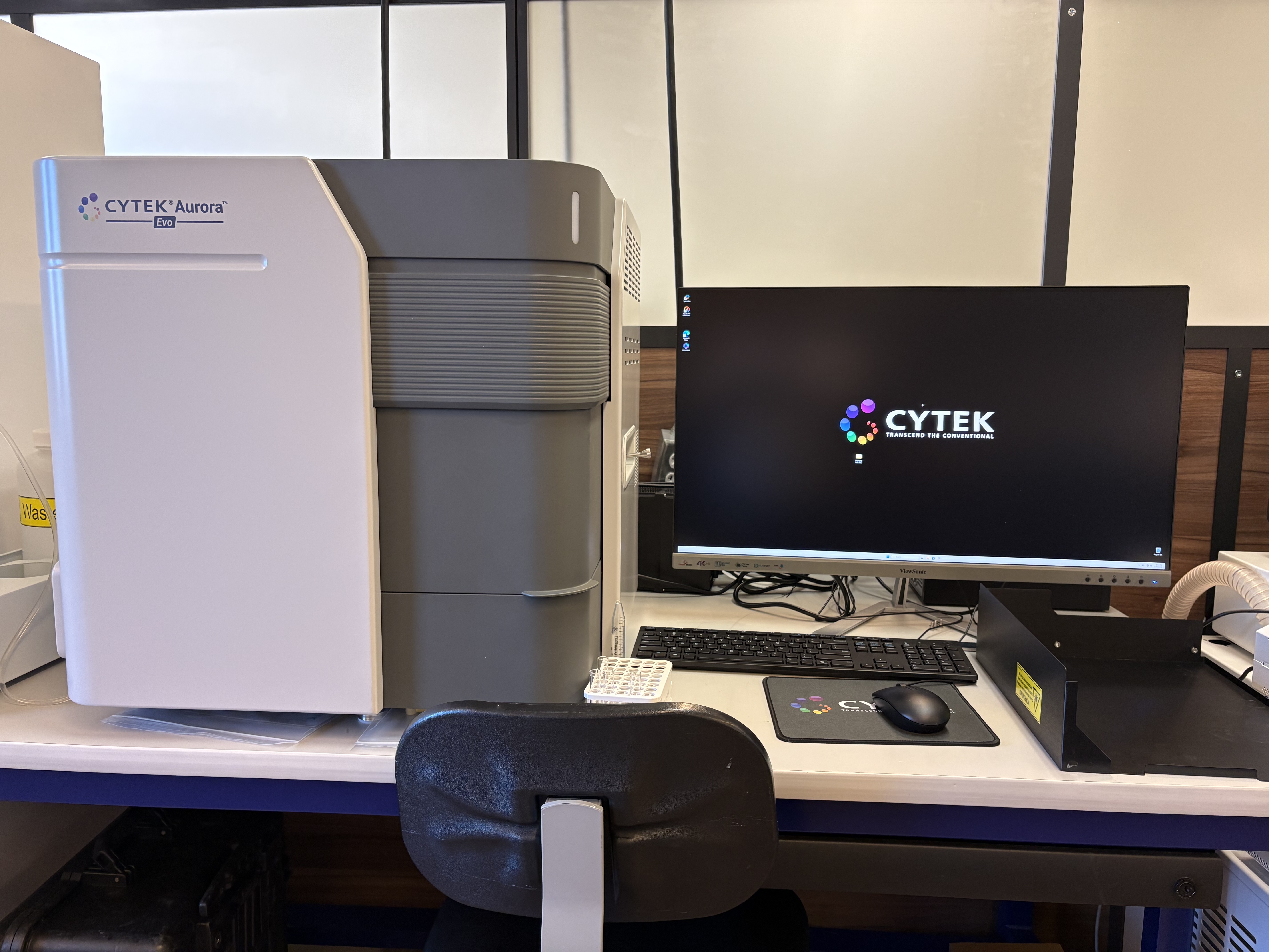 d 3-laser spectral flow cytometer designed for high-parameter single-cell immunophenotyping. Equipped with violet (405 nm), blue (488 nm), and red (640 nm) lasers, the system enables simultaneous measurement of 30–40+ fluorophore-conjugated markers through full-spectrum emission capture and computational spectral unmixing. This platform is ideally suited for comprehensive immune profiling in models of viral infection, inflammation, and tissue injury, supporting detailed characterization of lymphoid and myeloid subsets, activation states, and functional immune responses.
Agilent Biotek Cytation C10 Confocal Imager: The Agilent BioTek Cytation C10 is an 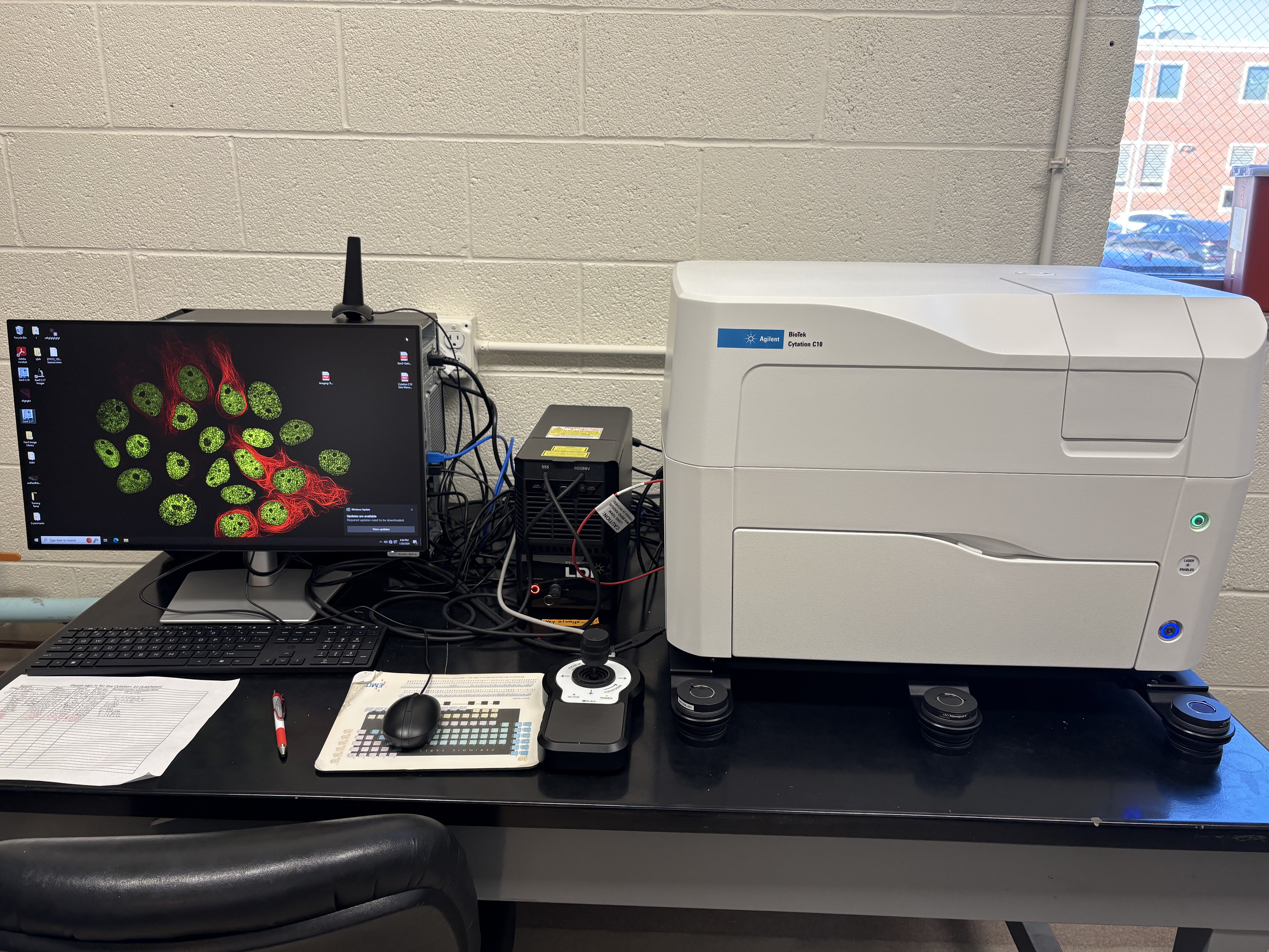 automated multimode imaging platform that combines confocal fluorescence microscopy with widefield brightfield, color brightfield, and phase-contrast imaging. It supports magnification from 1.25× to 60× (air) and 40× to 60× (water immersion). The system is equipped with a high-sensitivity Sony CMOS camera (16-bit grayscale) and a confocal spinning disk module with 60-micron pinholes for rapid, high-resolution optical sectioning. Cytation C10 includes environmental control with incubation up to 45°C and integrated shaking, enabling live-cell and kinetic imaging workflows. Data acquisition and analysis are performed using GEN5 software.
Bio-Rad Bio-Plex 200 system: 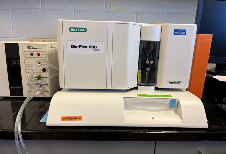 The Bio-Plex 200 system is a suspension array system which offers protein and nucleic acid researchers a reliable multiplex assay solution that permits analysis of up to 100 biomolecules in a single sample. The instrument is suitable to run any kit that is Luminex 200 compatible. For additional details see this SITE.
NanoCellect WOLF Cell Sorter: This Cell Sorter allows for gentle
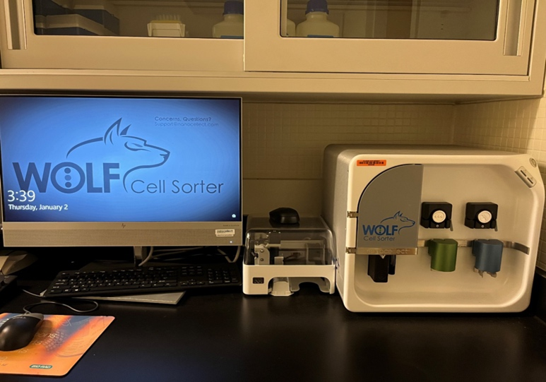 analysis, sorting and plating of cells based on its microfluidic technology. By using sterile, disposable fluidics, system clean-up becomes extremely fast and cross contaminations are prevented. Therefore, the WOLF Cell Sorter is intended for use in single-cell genomics, antibody production, and cell line development.
Olympus IX81 motorized fluorescence microscope: The IX81 is Olympus' most
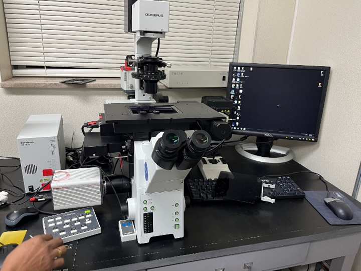 advanced motorized microscope, providing high standards of observation, measurement, and functional operation. A V-shaped optical path, improved fluorescence illuminators and further expanded UIS optics provide excellent performance for research applications.
BioTek Cytation 5: 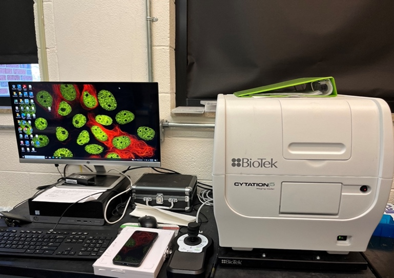 BioTek Cytation 5 combines automated microscopy and conventional microplate detection in a configurable, upgradable platform. The microscopy module offers up to 60x magnification in fluorescence, brightfield, high contrast brightfield, color brightfield, and phase contrast to address many applications and workflows. For application details, please see this SITE.
Zeiss LSM-910: The Zeiss LSM-910 is an advanced co 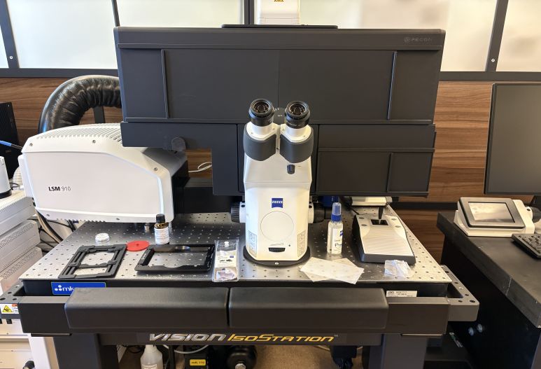 nfocal laser scanning microscope that integrates high-resolution fluorescence light microscopy with topography-based analysis to enable detailed characterization of complex 3D microstructures. This platform supports high-quality optical sectioning and three-dimensional reconstruction of biological specimens, providing exceptional spatial resolution and imaging depth. The instrument has broad applications in biomedical and translational research, including high-resolution imaging of cells and tissues, live-cell and time-lapse microscopy, visualization of pathogen–host interactions, subcellular localization of viral and immune markers, and quantitative 2D and 3D imaging of intact specimens. The Zeiss LSM-910 is a critical shared resource for studying cellular architecture, virus-host interaction, immune responses, and disease-associated tissue remodeling in infectious, inflammatory, and other models of diseases.
Molecular Devices SpectraMax M2: 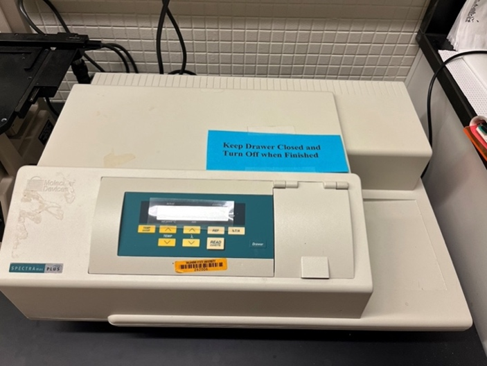 The SpectraMax M2 is a dual monochromator, multi-detection microwell reader with dual mode cuvette port and 96 or 384 microwell reader capability. It utilizes UV-Vis absorbance, as well as fluorescence intensity (FI) with optical performance comparable to top-level dedicated fluorescence spectrometers or spectrophotometers. Use this reader for Endpoint, Kinetic, Spectral, Area Well Scans, DNA, RNA and protein quantification and purity; PicoGreen, NanoOrange and Bradford; ELISA and enzyme kinetics; drug lysis profiles; live or dead viability, cytotoxicity, Caspase-3 and protease assays; cAMP assays using the CatchPoint kit.
Molecular Devices SpectraMax Plus: The SpectraMax Plus is a UV/Vis
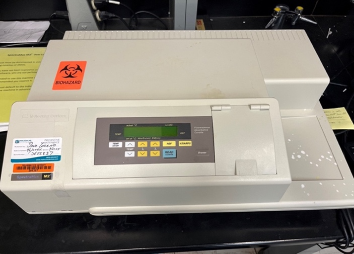 spectro-photometer/microwell reader with built-in cuvette port and microplate drawer. It can read any standard cuvette, micro cuvette, test tube and 96-well or UV-transparent microplate in the wavelength range of 190-1000 nm. It can read 96 wells, each well has its own sample beam and reference detector. Applications include DNA Analysis, ELISA, Endpoint Analysis, Kinetics.
ELISA Plate Washer: 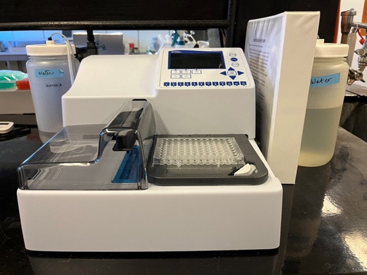 ELISA Plate Washer allows fast and easy washing of ELISA plates.
Leica CM1850: The Leica CM1850 is a high-precision cryostat microtome designed for sectioning frozen tissue
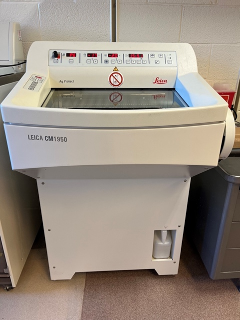 specimens in a variety of biological and medical applications. The CM1850 is widely used in research and clinical settings for creating thin, high-quality tissue sections that are used in various types of microscopy, including light, fluorescence, and electron microscopy.
Leica ASP300 Tissue Processor: The ASP300 performs a series of steps to
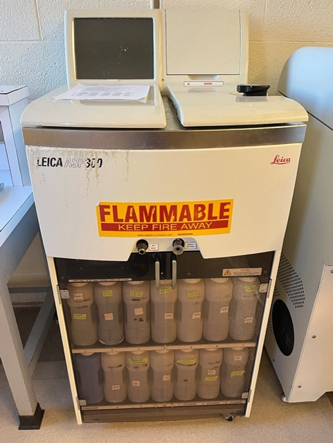 ensure that tissue samples are dehydrated, cleared and infiltrated with paraffin to allow for precise embedding and cutting. Tissues can then be used for a multitude of staining procedures.
Shandon Microtome and water bath: The microtome and wate
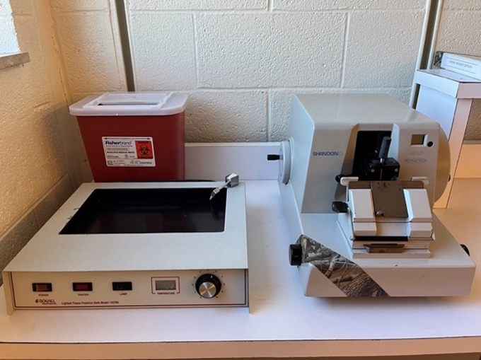 r bath will enable users to cut sections and mount on microscope slides. These are available for users on Do-It-Yourself basis and a small fee will be charged to equipment use. IPC staff will provide training to use this equipment.
The IPC Histology Lab can trim, process, embed, cut, and stain samples and slides using the following equipment on a fee-for-service basis. Equipment is managed, maintained and operated by IPC personnel.
Sakura Finetek Tissue-Tek VIP6 AI tissue processor: Sakura Finetek Tissue-Tek VIP6 AI tissue proc 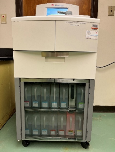 essor uses advanced infiltration designed to provide high-quality tissue processing results for all tissue types.
Leica EG1160 Embedding Station: Samples will be embedded
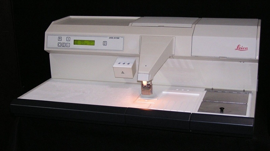 on a Leica EG1160 Embedding Station.
Leica HistoCore Biocut AS: 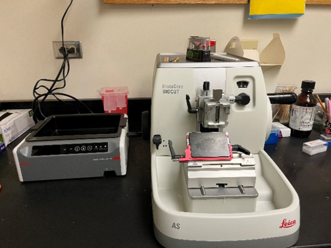 Leica HistoCore Biocut AS used to cut sections for investigators.
Sakura Finetek Tissue-Tek DRS 601 Slide Stainer  Slides are stained using the Sakura Finetek Tissue-Tek DRS 601 Slide Stainer.
OCRID Core Facilities User Satisfaction Survey
OCRID Core facilities are committed to providing top quality services and technical expertise in respiratory and infectious disease for center and non-center investigators. We encourage all users to fill out a simple, ten question survey. Your responses, comments, and suggestions can help us improve our services.